
Have you ever wondered how your heart knows to beat faster when you’re nervous about an AP exam? Or why a single scratch on your skin heals so perfectly that you can barely see where it was? The answer lies in one of biology’s most fascinating phenomena: cells talking to each other and knowing exactly when to divide. Welcome to AP Biology Unit 4: Cell Communication and Cell Cycle, where we’ll explore the intricate world of cell communication and the cell cycle.
This unit might seem daunting at first – after all, we’re diving into molecular conversations happening at microscopic levels. But here’s the thing: once you understand these processes, you’ll start seeing them everywhere in your daily life. From the insulin response after eating lunch to the way your immune system fights off that cold you caught from your lab partner, cellular communication is the foundation of everything that keeps you alive and functioning.
What You’ll Master by the End
By the time you finish this unit, you’ll have conquered these College Board learning objectives:
- ENE-3.A: Describe the mechanisms of cell communication through direct contact or chemical signaling
- ENE-3.B: Explain how cells respond to external and internal signals
- ENE-3.C: Describe the role of programmed cell death in development and maintenance of complex organisms
- IST-2.A: Describe the events that occur during the cell cycle
- IST-2.B: Explain how checkpoints in the cell cycle are regulated
- IST-2.C: Describe the characteristics and roles of cell cycle checkpoints in regulating the cell cycle
Don’t let these formal objectives intimidate you. Think of them as your roadmap to understanding some of the coolest processes in biology.
The Foundation: Why Cells Need to Communicate
Picture this: you’re in a group project with 37 trillion team members (that’s roughly how many cells you have). Now imagine trying to coordinate everyone without any communication system. Chaos, right? That’s exactly why cell communication evolved – it’s the biological equivalent of having everyone on the same group chat.
Cell communication, or cell signaling, is how cells coordinate their activities to maintain homeostasis, respond to environmental changes, and carry out complex biological processes. Without it, multicellular life as we know it simply wouldn’t exist. Your neurons couldn’t send signals to your muscles, your hormones couldn’t regulate your metabolism, and your immune system couldn’t protect you from pathogens.
Real-World Connection: When you smell fresh cookies baking, olfactory receptor cells in your nose detect odor molecules and send signals through your nervous system to your brain. Your brain then might signal your salivary glands to start producing saliva. This entire chain reaction happens because cells can communicate effectively.
Types of Cell Communication: From Neighbors to Long-Distance Relationships
Cell communication isn’t one-size-fits-all. Depending on the distance and type of signal, cells use different communication strategies:
Direct Contact Communication occurs when cells are physically touching. Think of it like whispering to the person sitting next to you in class. Gap junctions in animal cells and plasmodesmata in plant cells create direct channels between adjacent cells, allowing small molecules and ions to pass through. This type of communication is crucial for coordinating activities in tissues.
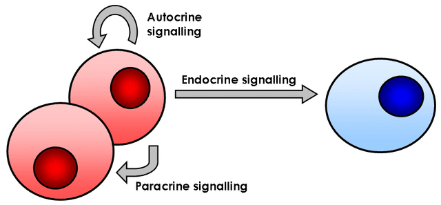
Local Signaling happens over short distances, typically through the release of chemical messengers called local regulators. Paracrine signaling affects nearby cells, while autocrine signaling affects the same cell that released the signal. It’s like speaking to everyone in your immediate vicinity.
Long-Distance Signaling involves hormones traveling through the circulatory system to reach target cells far from their origin. This is like sending a text message to someone across the country – the signal travels far but reaches its specific target.
Study Tip: Remember the three types using the acronym “DLL” – Direct, Local, Long-distance. Associate each with a real-world communication method to make it stick.
The Molecular Machinery: Signal Transduction Pathways
Now let’s dive into the nitty-gritty of how cells actually “talk” to each other. Signal transduction is essentially a cellular telephone system with three main components: reception, transduction, and response. Understanding this pathway is crucial for AP Biology success because it appears in multiple contexts throughout the exam.
Reception: The Cellular Doorbell
Reception occurs when a signaling molecule (ligand) binds to a receptor protein. This is like someone pressing your doorbell – the signal has arrived, and now your house (cell) knows someone’s there. Receptors can be located in different places:
Plasma Membrane Receptors are embedded in the cell membrane and detect water-soluble signals that can’t cross the lipid bilayer. Most hormones, neurotransmitters, and growth factors use this pathway. G protein-coupled receptors (GPCRs) are particularly important here – they’re involved in everything from your sense of smell to hormone regulation.
Intracellular Receptors are found inside the cell and detect lipid-soluble signals that can pass through the cell membrane. Steroid hormones like testosterone and estrogen use this pathway. These signals can directly affect gene expression by acting as transcription factors.
Quick Check: Can you predict whether insulin uses plasma membrane receptors or intracellular receptors? (Answer: Plasma membrane, because insulin is a protein hormone that can’t cross the lipid bilayer)
Transduction: The Cellular Game of Telephone
Once a signal is received, it must be converted into a form the cell can act upon. This is transduction – the process of converting one signal into another. Think of it like translating a message from one language to another, except cells often play an elaborate game of telephone where the message gets passed through multiple intermediaries.
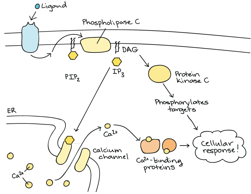
Protein Phosphorylation is the most common mechanism of transduction. When a protein gets phosphorylated (has a phosphate group added), it typically changes shape and activity. Kinases add phosphate groups, while phosphatases remove them. This creates an on/off switch that can be rapidly regulated.
Second Messengers amplify the original signal. Common second messengers include cyclic AMP (cAMP), cyclic GMP (cGMP), and calcium ions. A single hormone molecule binding to a receptor might trigger the production of thousands of second messenger molecules, creating a powerful amplification effect.
Cascade Effects occur when the activation of one enzyme leads to the activation of many more enzymes in a chain reaction. The MAPK (mitogen-activated protein kinase) pathway is a classic example that’s frequently tested on the AP exam.
Common Mistake Alert: Students often confuse the terms “ligand” and “receptor.” Remember: the ligand is the key (signaling molecule), and the receptor is the lock (protein that binds the ligand).
Response: When Cells Take Action
The final stage of signal transduction is the cellular response. This could involve:
- Activation or inhibition of enzymes – changing metabolic pathways
- Changes in gene expression – turning genes on or off
- Alterations in cell shape or movement – important for development and immune responses
- Modification of membrane permeability – affecting what enters or leaves the cell
The beauty of signal transduction is its specificity and amplification. One hormone molecule can trigger responses in millions of target cells, each responding according to its specific receptor complement and cellular machinery.
Hormones and Cell Signaling: Your Body’s Chemical Messengers
Hormones are probably the most familiar example of cell communication, and they’re heavily featured on the AP Biology exam. Understanding how different hormones work provides excellent examples of signal transduction principles in action.
Insulin: The Master Metabolic Regulator
Let’s use insulin as our detailed example because it beautifully illustrates signal transduction principles and connects to multiple other AP Biology topics.
When you eat a slice of pizza, your blood glucose levels rise. Beta cells in your pancreas detect this increase and release insulin into your bloodstream. Insulin then travels to target cells throughout your body, particularly in muscle, fat, and liver tissue.
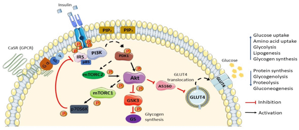
Insulin binds to insulin receptors on the surface of target cells. These receptors are receptor tyrosine kinases (RTKs), which undergo autophosphorylation when insulin binds. This phosphorylation triggers a cascade of intracellular events:
- The phosphorylated receptor activates insulin receptor substrates (IRS proteins)
- IRS proteins activate PI3K (phosphoinositide 3-kinase)
- PI3K produces PIP3, which activates protein kinase B (PKB/Akt)
- PKB/Akt triggers multiple responses including glucose uptake and glycogen synthesis
Real-World Connection: Understanding insulin signaling is crucial for comprehending diabetes. In Type 1 diabetes, beta cells can’t produce insulin. In Type 2 diabetes, cells become resistant to insulin signaling, often due to problems in the signal transduction pathway.
Epinephrine: The Fight-or-Flight Hormone
Epinephrine (adrenaline) provides an excellent contrast to insulin and showcases G protein-coupled receptor signaling. When you’re startled by a sudden loud noise, your adrenal glands release epinephrine.
Epinephrine binds to G protein-coupled receptors on target cells. This activates a G protein, which then activates adenylyl cyclase. Adenylyl cyclase converts ATP to cyclic AMP (cAMP), a crucial second messenger. cAMP then activates protein kinase A (PKA), which phosphorylates multiple target proteins.
The result? Your heart rate increases, your pupils dilate, and your liver releases glucose into your bloodstream – all preparing your body for action.
Study Tip: Create a comparison table of different hormones, their receptor types, second messengers, and primary effects. This will help you recognize patterns and predict outcomes on exam questions.
Apoptosis: Programmed Cell Death
This might sound morbid, but programmed cell death (apoptosis) is actually essential for life. It’s one of those counterintuitive biological concepts that frequently appears on AP exams because it challenges students’ assumptions about what’s “good” for organisms.
Why Cells Need to Die
Apoptosis serves several crucial functions:
Development: During embryonic development, apoptosis removes unnecessary cells. The webbing between your fingers and toes disappears through apoptosis. Without it, you’d have duck-like appendages!
Homeostasis: In adult organisms, apoptosis maintains proper cell numbers. Your body produces about 25 million new cells every second, so an equal number must die to maintain balance.
Protection: Cells that are damaged, infected, or potentially cancerous undergo apoptosis to protect the organism. It’s like a cellular suicide mission for the greater good.
The Apoptotic Pathway
Apoptosis can be triggered by internal or external signals. Internal triggers include DNA damage, cellular stress, or developmental cues. External triggers include cytotoxic T cells or growth factor withdrawal.
The process involves a family of proteins called caspases (cysteine-aspartate proteases). These enzymes are normally inactive but become activated during apoptosis. Once activated, they systematically dismantle the cell:
- Initiator caspases respond to apoptotic signals
- Executioner caspases are activated by initiator caspases
- The cell shrinks and fragments its DNA
- The cell breaks into apoptotic bodies
- Phagocytes engulf and digest the apoptotic bodies
Quick Check: What’s the difference between apoptosis and necrosis? (Answer: Apoptosis is programmed cell death that’s beneficial to the organism, while necrosis is accidental cell death due to injury or disease)
The Cell Cycle: Life’s Most Important Schedule
Now we shift gears from cell communication to cell division. The cell cycle is the highly regulated process by which cells grow and divide to produce two daughter cells. Understanding this process is fundamental to biology because cell division is how organisms grow, repair tissues, and reproduce.
The Phases: A Cellular Journey
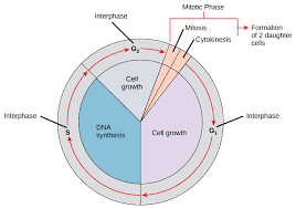
The cell cycle consists of distinct phases, each with specific activities and checkpoints:
G1 Phase (Gap 1): This is the growth phase where cells increase in size and synthesize enzymes and proteins needed for DNA replication. Most of your body’s cells spend the majority of their time in G1. Some cells exit the cell cycle and enter G0, a quiescent state where they don’t prepare for division.
S Phase (Synthesis): DNA replication occurs during this phase. Each chromosome is duplicated, creating sister chromatids joined at the centromere. This is perhaps the most critical phase because errors here can lead to mutations.
G2 Phase (Gap 2): Cells continue growing and produce proteins necessary for chromosome condensation and mitosis. The cell also checks that DNA replication was completed successfully.
M Phase (Mitosis): The cell divides, distributing one copy of each chromosome to each daughter cell. This phase includes both nuclear division (mitosis proper) and cytoplasmic division (cytokinesis).
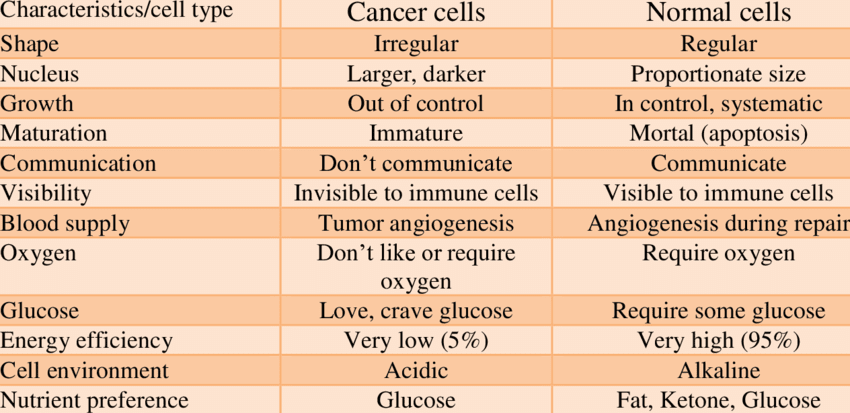
Real-World Connection: Cancer cells have disrupted cell cycles – they’ve lost the ability to respond to normal stop signals and divide uncontrollably. Understanding normal cell cycle regulation helps us comprehend how cancer develops and how treatments work.
Cell Cycle Duration: Not All Cells Are Equal
Different cell types have vastly different cell cycle durations. Embryonic cells might complete a cycle in 30 minutes, while adult liver cells might take a year or more. Some neurons never divide after maturation, remaining permanently in G0.
This variation reflects the different roles cells play in the organism. Rapidly dividing cells like those in your intestinal lining need quick turnover to replace damaged cells, while neurons need stability to maintain complex connections.
Cell Cycle Checkpoints: Quality Control Systems
One of the most important concepts in cell cycle regulation is the checkpoint system. These are molecular mechanisms that ensure each phase of the cell cycle is completed correctly before proceeding to the next phase. Think of them as quality control inspectors on an assembly line.
The G1/S Checkpoint (Restriction Point)
This checkpoint occurs near the end of G1 and determines whether the cell will proceed with DNA replication. The cell assesses:
- Cell size: Is the cell large enough to divide successfully?
- Nutrients: Are sufficient nutrients available for DNA synthesis?
- Growth factors: Are external signals promoting cell division present?
- DNA damage: Is the DNA intact and undamaged?
The p53 protein plays a crucial role here. Often called the “guardian of the genome,” p53 detects DNA damage and can halt the cell cycle, activate DNA repair mechanisms, or trigger apoptosis if damage is too severe.
Study Tip: Remember p53 as the cellular “emergency brake” that stops division when problems are detected.
The Intra-S Phase Checkpoint
This checkpoint monitors DNA replication during S phase. If replication forks stall or DNA damage occurs during synthesis, this checkpoint activates repair mechanisms and may slow down or halt replication until problems are resolved.
The G2/M Checkpoint
Before entering mitosis, cells must verify that DNA replication is complete and that there’s no DNA damage. This checkpoint ensures that each daughter cell will receive a complete, undamaged set of chromosomes.
The Spindle Checkpoint (M Checkpoint)
During mitosis, this checkpoint ensures that all chromosomes are properly attached to spindle fibers before allowing anaphase to proceed. This prevents chromosome loss or unequal distribution to daughter cells.
Cyclins and CDKs: The Cell Cycle’s Molecular Clock
The cell cycle is regulated by a sophisticated molecular machinery involving cyclins and cyclin-dependent kinases (CDKs). This system ensures that cell cycle events occur in the correct order and at the appropriate times.
How the System Works
Cyclins are proteins whose concentrations rise and fall throughout the cell cycle. They’re named “cyclins” because their levels cycle up and down. Different cyclins are active during different phases:
- G1/S cyclins promote entry into S phase
- S cyclins are required for DNA replication
- M cyclins drive entry into mitosis
Cyclin-dependent kinases (CDKs) are enzymes that become active only when bound to the appropriate cyclin. Once activated, CDK-cyclin complexes phosphorylate target proteins to drive cell cycle progression.
The system works like a molecular clock:
- Cyclin levels rise during specific phases
- Cyclins bind to and activate CDKs
- Active CDK-cyclin complexes phosphorylate target proteins
- These phosphorylation events drive cell cycle progression
- Cyclins are degraded, inactivating CDKs
- The cycle repeats
Common Mistake Alert: Students often think CDKs vary in concentration throughout the cell cycle. In reality, CDK levels remain relatively constant – it’s the cyclin levels that oscillate.
Regulation of CDK Activity
CDK activity is controlled through multiple mechanisms:
Cyclin binding: CDKs are only active when bound to appropriate cyclins
Phosphorylation: CDKs can be phosphorylated at activating or inhibiting sites
CDK inhibitors (CKIs): These proteins bind to and inactivate CDK-cyclin complexes
Targeted degradation: Cyclins are marked for destruction by the ubiquitin-proteasome system
This multi-layered regulation ensures precise control over cell cycle progression and provides multiple points where the cycle can be halted if problems arise.
Growth Factors and External Regulation
While internal mechanisms like cyclins and CDKs drive the cell cycle, external signals significantly influence whether cells divide. Growth factors are proteins that promote cell division, while growth inhibitors prevent it.
Positive Regulators: Growth Factors
Platelet-derived growth factor (PDGF) stimulates cell division in response to tissue damage. When you get a cut, platelets release PDGF to promote healing through increased cell division.
Epidermal growth factor (EGF) stimulates division of skin cells and other epithelial tissues.
Insulin-like growth factors (IGFs) promote general body growth and cell division throughout development.
These factors typically work by binding to receptor tyrosine kinases, activating signal transduction pathways that ultimately promote cyclin production and CDK activation.
Negative Regulators: Growth Inhibitors
Contact inhibition occurs when cells stop dividing upon contacting neighboring cells. This prevents overcrowding and maintains proper tissue architecture.
Density-dependent inhibition causes cells to stop dividing when they reach a certain density, even without direct contact.
Growth inhibitory factors like TGF-β (transforming growth factor beta) actively suppress cell division in many cell types.
Real-World Connection: Cancer cells often lose responsiveness to growth inhibitors, which contributes to their uncontrolled division. Understanding normal growth regulation helps explain how cancer develops and suggests therapeutic targets.
Cancer: When Cell Cycle Control Goes Wrong
Cancer represents a breakdown in normal cell cycle control. Understanding cancer through the lens of cell cycle regulation provides excellent exam preparation because it integrates multiple concepts from this unit.
The Hallmarks of Cancer Cells
Cancer cells differ from normal cells in several key ways:
Ignore growth inhibitory signals: They don’t respond to contact inhibition or growth inhibitory factors
Don’t require growth factors: They can divide without external growth signals
Avoid apoptosis: They resist programmed cell death even when damaged
Replicate indefinitely: They bypass normal limits on cell division
Invade and metastasize: They can spread to other parts of the body
Oncogenes and Tumor Suppressors
Cancer typically results from mutations in two types of genes:
Oncogenes are mutated versions of normal genes (proto-oncogenes) that promote cell division. When oncogenes are overactive, they drive excessive cell division. Examples include mutated growth factor receptors or cell cycle regulatory proteins.
Tumor suppressor genes normally prevent uncontrolled cell division. When these genes are lost or inactivated, cells lose important brakes on division. p53 and Rb (retinoblastoma protein) are classic examples.
The Two-Hit Hypothesis explains why cancer typically requires multiple mutations. Most tumor suppressor genes follow this pattern – you need to lose both copies (one from each parent) for cancer to develop.
Study Tip: Think of oncogenes as a stuck accelerator pedal (promoting division) and tumor suppressors as broken brakes (failing to stop division). Cancer occurs when you have both problems simultaneously.
Connecting Cell Communication and Cell Cycle
These two major topics aren’t separate – they’re intimately connected. Cell communication pathways frequently regulate cell cycle progression, creating an integrated system that maintains tissue homeostasis.
Growth Factor Signaling and Cell Cycle Entry
Growth factors like PDGF bind to receptor tyrosine kinases, triggering signal transduction cascades that ultimately increase cyclin production. This connection explains how external signals can promote cell division when needed (like during wound healing) and suppress it when appropriate (like in mature tissues).
p53: The Master Integrator
The p53 protein beautifully illustrates the connection between cell communication and cell cycle control. p53 responds to various stress signals (DNA damage, hypoxia, oncogene activation) and can halt the cell cycle, promote DNA repair, or trigger apoptosis. It’s essentially a communication hub that integrates multiple signals to make cell fate decisions.
Contact-Dependent Signaling
When cells contact each other, they exchange signals through surface receptors that can inhibit cell division. This contact inhibition is crucial for maintaining proper tissue organization and preventing tumor formation.
Practice Questions: Test Your Understanding
Let’s put your knowledge to the test with some AP-style questions. Remember, the AP Biology exam includes both multiple choice and free response questions, so practicing both formats is essential.
Multiple Choice Questions
Question 1: A researcher treats cells with a chemical that prevents cyclin degradation. What would be the most likely result?
A) Cells would be unable to enter S phase
B) Cells would arrest at the G2/M checkpoint
C) Cells would progress through the cell cycle too rapidly
D) Cells would immediately undergo apoptosis
Answer: C. If cyclins aren’t degraded, CDKs would remain active longer than normal, driving continuous cell cycle progression.
Question 2: Which of the following best describes the role of second messengers in signal transduction?
A) They directly bind to DNA to alter gene expression
B) They amplify the original signal within the cell
C) They transport signals between different cells
D) They degrade signaling molecules to terminate responses
Answer: B. Second messengers like cAMP amplify signals by being produced in large quantities in response to a single hormone binding event.
Question 3: A mutation in the p53 gene would most likely result in:
A) Increased sensitivity to growth factors
B) Enhanced DNA repair capabilities
C) Reduced ability to detect DNA damage
D) Faster progression through S phase
Answer: C. p53 is crucial for detecting DNA damage and halting the cell cycle when problems occur.
Free Response Practice
Question: Describe the process of signal transduction using a specific hormone as an example. Include the following in your answer:
- The three stages of signal transduction
- The role of receptors and second messengers
- How the signal is amplified
- The ultimate cellular response
Sample Answer Framework:
Begin with insulin as your example. Describe reception (insulin binding to receptor tyrosine kinases), transduction (autophosphorylation leading to activation of downstream signaling cascades), and response (increased glucose uptake and glycogen synthesis). Explain amplification through enzyme cascades where one activated enzyme activates many others. Conclude with the physiological significance of this response in glucose homeostasis.
Data Analysis Question
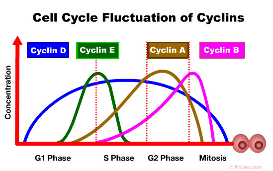
Question: The graph above shows the concentration of different cyclins throughout the cell cycle.
a) Identify which cyclin would be most active during DNA replication and explain your reasoning.
b) Predict what would happen if M cyclin levels remained high after mitosis was complete.
c) Explain how this data supports the role of cyclins in cell cycle regulation.
Answer Guide:
a) S cyclin would be most active during DNA replication because its peak concentration corresponds to S phase when DNA synthesis occurs.
b) High M cyclin levels would prevent cells from exiting mitosis properly, potentially leading to cell cycle arrest or abnormal cell division.
c) The cyclical nature of cyclin concentrations provides evidence that these proteins regulate cell cycle timing by being present only when their specific functions are needed.
Common Exam Mistakes and How to Avoid Them
Based on years of AP Biology exams, certain mistakes appear repeatedly. Here’s how to avoid them:
Confusing Growth Factors with Hormones: Remember that all growth factors are signaling molecules, but not all hormones are growth factors. Growth factors specifically promote cell division.
Mixing Up Cyclin and CDK Functions: Cyclins vary in concentration (they cycle), while CDKs remain relatively constant but are activated by cyclin binding.
Forgetting Checkpoint Functions: Each checkpoint has a specific purpose. Don’t just memorize their names – understand what they check for and why that’s important.
Oversimplifying Signal Transduction: Signal transduction isn’t just “hormone binds, cell responds.” Make sure you understand the intermediate steps and why amplification is crucial.
Ignoring the Connection Between Topics: Cell communication and cell cycle aren’t separate units. They work together to maintain homeostasis and coordinate cellular activities.
Real-World Applications: Where This Knowledge Matters
Understanding cell communication and the cell cycle isn’t just academic – it has profound real-world applications:
Medical Research: Drug development often targets specific steps in signal transduction pathways. Many cancer treatments work by disrupting cell cycle progression or inducing apoptosis in cancer cells.
Biotechnology: Scientists manipulate cell signaling pathways to produce insulin in bacteria, grow tissues in culture, and develop new therapeutic approaches.
Agricultural Science: Understanding how plant hormones communicate growth and development information helps develop better crop varieties and growth regulators.
Environmental Science: Many environmental toxins work by disrupting normal cell communication or cell cycle control, leading to cancer or developmental abnormalities.
Memory Techniques for Complex Pathways
Cell communication and cell cycle involve many detailed pathways. Here are some memory techniques that work:
For Signal Transduction: Remember “ReTaRd” (Reception, Transduction, Response) for the three stages.
For Cell Cycle Phases: “Go Sit Go More” (G1, S, G2, M) helps remember the order.
For Checkpoint Functions: Create stories or acronyms for what each checkpoint checks. For example, the G1/S checkpoint is like a “security checkpoint” that checks for damage before allowing DNA replication.
For Hormone Examples: Group similar hormones together and note their common features (peptide vs. steroid, plasma membrane vs. intracellular receptors).
Looking Ahead: Connections to Other Units
Unit 4 doesn’t exist in isolation. Here’s how it connects to other AP Biology units:
Unit 2 (Cell Structure and Function): Cell communication builds on membrane structure and transport mechanisms.
Unit 3 (Cellular Energetics): Many signaling pathways regulate metabolic processes you learned in Unit 3.
Unit 5 (Heredity): Cell cycle regulation is crucial for understanding how genetic information is passed to daughter cells.
Unit 6 (Gene Expression): Many cellular responses involve changes in gene expression.
Unit 7 (Natural Selection): Cell communication systems evolved to help organisms survive and reproduce.
Unit 8 (Ecology): Population growth often depends on cell division rates and responses to environmental signals.
Your Path to AP Success
Mastering AP Biology Unit 4 requires more than memorization – it requires understanding the elegant connections between cellular communication and division. These processes represent some of biology’s most sophisticated regulatory mechanisms, evolved over millions of years to maintain the delicate balance necessary for life.
As you continue your AP Biology journey, remember that these concepts aren’t just test material – they’re the foundation for understanding how life works at its most fundamental level. Every time your heart beats, every time a cut heals, every time your body responds to stress, you’re witnessing the cellular communication and division processes you’ve studied in this unit.
The key to success on the AP exam is understanding these concepts deeply enough to apply them to new situations. Don’t just memorize signal transduction pathways – understand why they work the way they do. Don’t just know the cell cycle phases – comprehend how their regulation maintains organism health.
With consistent study, active practice, and a focus on understanding rather than memorization, you’ll not only succeed on the AP Biology exam but also gain a deeper appreciation for the incredible complexity and beauty of life at the cellular level. The molecular conversations happening in your cells right now as you read this are more sophisticated than any human communication system – and now you understand how they work.
Remember, every AP Biology student who has succeeded before you has mastered these same concepts. With dedication and the right study strategies, you can join their ranks and earn the college credit you’re working toward. The cellular world is waiting for you to explore it – dive in and discover the amazing processes that make life possible.
This comprehensive guide covers all essential aspects of AP Biology Unit 4. Use it as your primary study resource, supplemented with practice problems from your textbook and released AP exams. Good luck with your studies, and remember – understanding these concepts now will serve you well in college biology courses and beyond.
Read Also –
2 thoughts on “Mastering AP Biology Unit 4: Cell Communication and Cell Cycle – Your Complete Guide to Exam Success”