
Have you ever wondered why you can survive for weeks without food but only days without water? The answer lies in the microscopic world of Cells – the fundamental units of life that make up every living organism on Earth. From the single-celled bacteria in your gut that help digest food to the trillions of specialized cells that make up your body, understanding cellular biology isn’t just academic knowledge – it’s the key to understanding life itself.
As an AP Biology student, Unit 2: Cells represents one of the most crucial foundations you’ll build throughout the course. This unit doesn’t just teach you about tiny structures you can’t see; it explains how every biological process you’ll encounter in later units actually works at the molecular level. Whether you’re struggling to understand membrane transport or feeling overwhelmed by cellular respiration, this comprehensive guide will transform complex cellular concepts into clear, manageable knowledge that sticks.
Learning Objectives: What You Need to Master
By the end of this unit, you should confidently demonstrate understanding of these College Board learning objectives:
- ENE-1.A: Explain how the structure of cell membranes relates to selective permeability
- ENE-1.B: Describe the roles of each of the components of the cell membrane
- ENE-1.C: Explain how concentration gradients affect the movement of molecules across membranes
- ENE-1.D: Describe the processes that allow ions and other molecules to move across membranes
- ENE-1.E: Describe the membrane-bound structures of the eukaryotic cell
- ENE-1.F: Explain how subcellular components and organelles contribute to the function of the cell
- ENE-1.G: Describe the structural features of a prokaryotic cell
- ENE-1.H: Explain how internal membranes and membrane-bound organelles contribute to compartmentalization of eukaryotic cell functions
Chapter 1: The Cell Theory and Why Size Matters
Let’s start with a story. In 1665, Robert Hooke was examining a thin slice of cork under his primitive microscope when he noticed tiny, box-like structures that reminded him of monastery cells. He had no idea he was looking at the building blocks of all life on Earth. This observation, along with work by Schleiden, Schwann, and Virchow, led to the cell theory – one of biology’s most fundamental principles.
The cell theory states three key points that you absolutely must know for the AP exam:
- All living things are composed of one or more cells
- The cell is the basic unit of life
- All cells arise from existing cells
But here’s what many students miss: understanding why cells are the size they are is crucial for grasping cellular transport and energy processes. Think about why elephants don’t have cells the size of basketballs, or why bacteria stay microscopic.
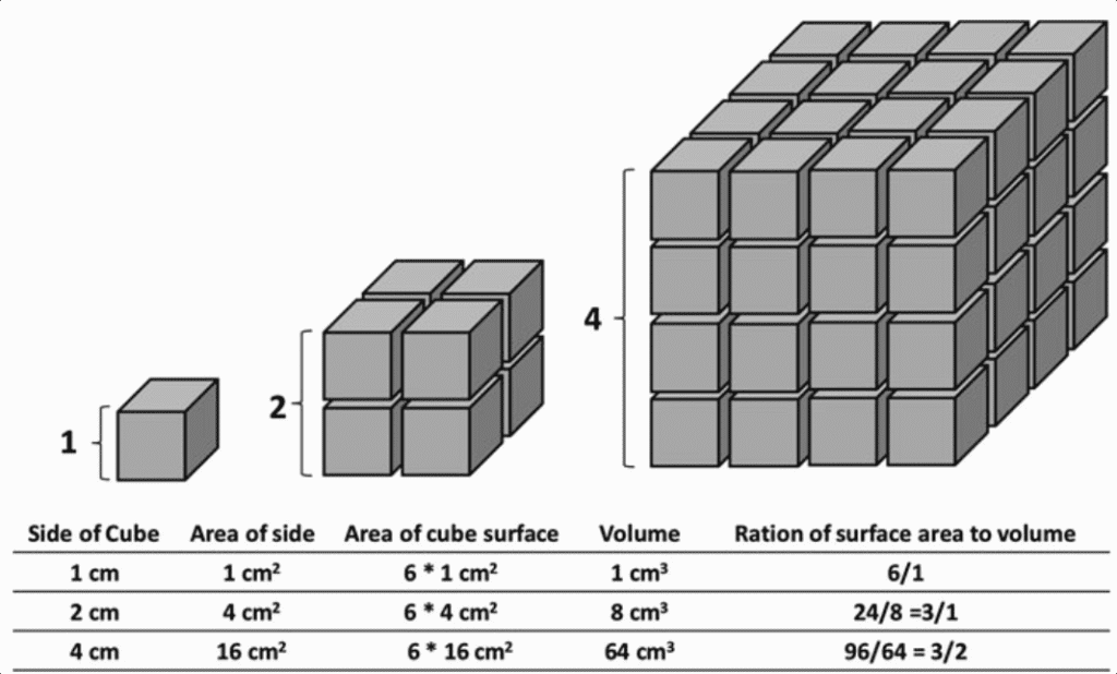
The surface area-to-volume ratio explains everything. As a cell grows larger, its volume increases much faster than its surface area. Since the cell membrane is where all the action happens – nutrients coming in, wastes going out, chemical signals being received – a cell that’s too large can’t efficiently exchange materials with its environment.
Study Tip: Remember the SA:V ratio with this analogy: imagine trying to feed a classroom of 30 students through a single doorway versus feeding 3 students through the same doorway. The bigger the “classroom” (cell volume), the more inadequate that single “doorway” (surface area) becomes.
Key Takeaways:
- Cell theory explains the fundamental nature of all living organisms
- Surface area-to-volume ratio limits cell size and explains cellular efficiency
- Small cells are more efficient at exchanging materials with their environment
Chapter 2: Prokaryotic Cells – The Minimalists of Life
Before we dive into the complexity of eukaryotic cells, let’s appreciate the elegant simplicity of prokaryotes. These cells – bacteria and archaea – have been thriving on Earth for about 3.8 billion years, and they’ve mastered survival with remarkable efficiency.
When I first started teaching AP Biology, students often dismissed prokaryotes as “simple” or “primitive.” This couldn’t be further from the truth. Prokaryotes are incredibly sophisticated; they’re just organized differently than eukaryotic cells.
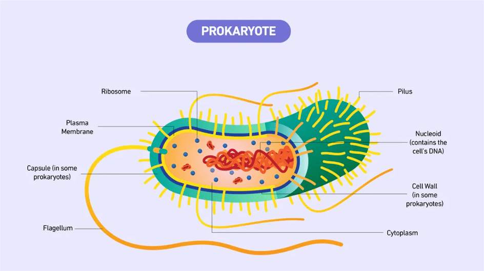
Essential Prokaryotic Structures:
Cell Wall: Think of this as the cell’s suit of armor. In bacteria, it’s made primarily of peptidoglycan, a complex polymer that provides structural support and protection. This is why antibiotics like penicillin work – they target peptidoglycan synthesis, causing bacterial cell walls to weaken.
Cell Membrane: Despite lacking internal membranes, prokaryotes have highly specialized cell membranes that perform many functions that eukaryotes delegate to organelles. The membrane contains transport proteins, electron transport chains for energy production, and sensors for environmental changes.
Nucleoid Region: This isn’t a true nucleus because it lacks a membrane, but it’s where the prokaryotic chromosome resides. The single, circular chromosome is supercoiled and associated with proteins to fit into this compact space.
Ribosomes: Prokaryotic ribosomes (70S) are smaller than eukaryotic ribosomes (80S) and have slightly different structures. This difference is why some antibiotics can target bacterial ribosomes without affecting human ribosomes.
Real-World Connection: The current COVID-19 pandemic has reminded us that while viruses aren’t cells, understanding prokaryotic structure helps us appreciate why bacteria and viruses require different treatment approaches. Antibiotics work on bacterial cell structures but are useless against viruses.
Common Mistake Alert: Many students confuse prokaryotic and eukaryotic ribosomes. Remember: prokaryotes have 70S ribosomes (made of 30S and 50S subunits), while eukaryotes have 80S ribosomes (made of 40S and 60S subunits). The ‘S’ stands for Svedberg units, which measure sedimentation rate, not size.
Key Takeaways:
- Prokaryotes lack membrane-bound organelles but are highly efficient
- Cell wall composition varies between bacteria and archaea
- Prokaryotic ribosomes differ from eukaryotic ribosomes in size and structure
- The nucleoid region contains the prokaryotic chromosome
Chapter 3: Eukaryotic Cells – The Specialists
If prokaryotes are minimalists, eukaryotes are the ultimate specialists. The evolution of membrane-bound organelles allowed eukaryotic cells to compartmentalize functions, leading to incredible diversity and complexity. From the neurons in your brain to the chloroplasts in a leaf, eukaryotic cells have revolutionized life on Earth.
The key innovation that defines eukaryotes is compartmentalization. Imagine trying to cook a five-course meal in a studio apartment versus a kitchen with separate prep areas, ovens, and storage spaces. Eukaryotic cells are like that well-designed kitchen – each organelle has a specific function and optimal environment.
The Nucleus: Command Center
The nucleus isn’t just where DNA hangs out; it’s a sophisticated control center that regulates gene expression and coordinates cellular activities. The nuclear envelope, with its nuclear pores, acts like a selective border control, allowing specific molecules in and out while maintaining the unique environment inside.
Nuclear Pores: These aren’t simple holes. They’re complex protein structures that actively transport materials. Small molecules can diffuse through, but larger molecules like mRNA and proteins require specific transport mechanisms.
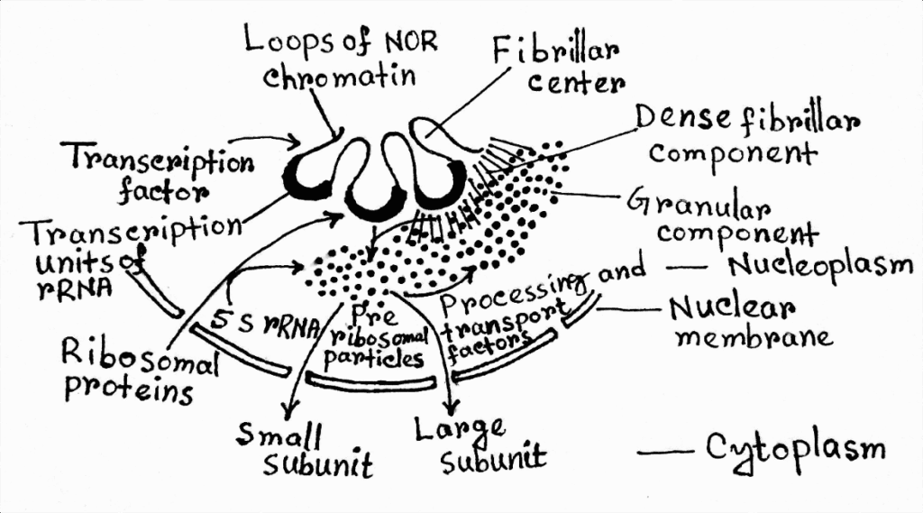
Nucleolus: This dense region within the nucleus is where ribosomal RNA is synthesized and ribosomal subunits are assembled. It’s not membrane-bound but represents a distinct functional compartment.
The Endomembrane System: Cellular Assembly Line
One of the most challenging concepts for AP Biology students is understanding how the endomembrane system works as an integrated unit. Think of it as a cellular assembly line where proteins and lipids are manufactured, modified, packaged, and shipped to their destinations.
Endoplasmic Reticulum (ER):
- Rough ER: Studded with ribosomes, this is where proteins destined for secretion, membrane incorporation, or organelle targeting are synthesized. The ribosomes translate mRNA while threading the growing protein into the ER lumen.
- Smooth ER: Lacks ribosomes and specializes in lipid synthesis, steroid hormone production, and detoxification. In liver cells, the smooth ER is extensive due to its role in detoxifying harmful substances.
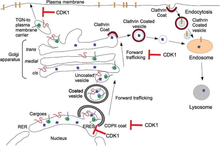
Golgi Apparatus: Often called the “post office” of the cell, the Golgi receives proteins from the ER, modifies them (adding carbohydrates, for example), and packages them for transport to their final destinations.
Study Tip: To remember the endomembrane system pathway, use this sequence: “Really Good Students Always Pass Tests” (Ribosome → Golgi → Sorting → Appropriate destination → Purpose fulfilled → Target reached).
Lysosomes: Cellular Recycling Centers
Lysosomes are fascinating organelles that demonstrate the importance of pH in cellular function. These membrane-bound sacs maintain an acidic environment (pH ~4.5) that activates their digestive enzymes. This acidic environment also protects the cell – if a lysosome accidentally breaks, the enzymes become inactive in the neutral cytoplasm.
Real-World Connection: Lysosomal storage diseases like Tay-Sachs demonstrate what happens when these organelles malfunction. Understanding lysosomes helps medical researchers develop treatments for these genetic disorders.
Mitochondria: The Powerhouses
Every AP Biology student learns that mitochondria are the “powerhouses of the cell,” but understanding why requires grasping their unique structure and evolutionary origin. Mitochondria have their own DNA, ribosomes, and can reproduce independently – evidence supporting the endosymbiotic theory.
Double Membrane Structure:
- Outer membrane: Permeable to small molecules
- Inner membrane: Highly folded into cristae, impermeable to most substances
- Intermembrane space: Where protons accumulate during ATP synthesis
- Matrix: Where the citric acid cycle occurs
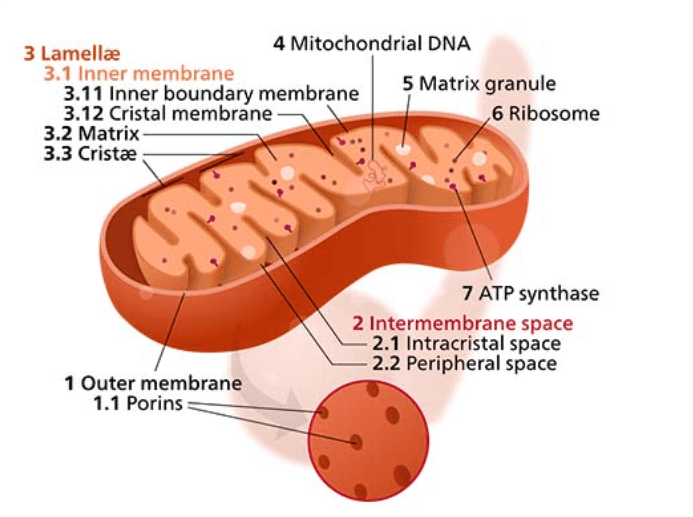
The folded inner membrane (cristae) dramatically increases surface area for ATP synthesis. Think of it like having more lanes on a highway – more space means more efficient traffic flow.
Key Takeaways:
- Eukaryotic cells use compartmentalization for efficiency
- The endomembrane system works as an integrated manufacturing and shipping network
- Mitochondrial structure directly relates to function in ATP production
- Each organelle has evolved specific features for its role
Chapter 4: Plant Cells – Green Machines
Plant cells share all the organelles found in animal cells but have three additional structures that make photosynthesis and structural support possible: chloroplasts, a large central vacuole, and a cell wall.
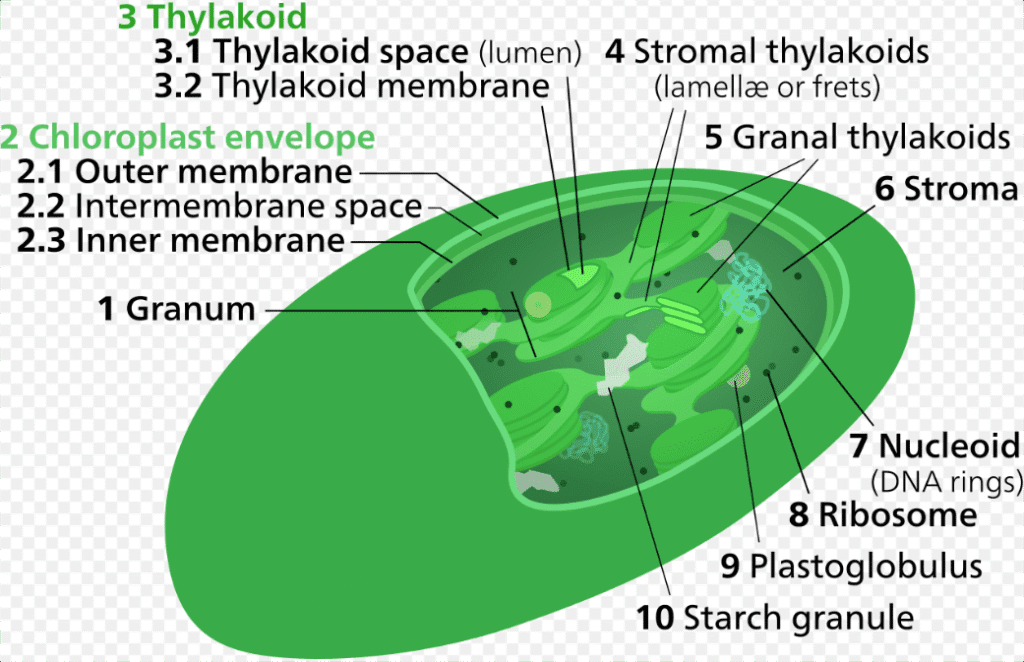
Chloroplasts: Solar Power Plants
Like mitochondria, chloroplasts have a double membrane and their own DNA, supporting the endosymbiotic theory. But chloroplasts have an additional internal membrane system – the thylakoids – where photosynthesis occurs.
Thylakoid Structure:
- Thylakoid membranes: Where light reactions occur
- Thylakoid lumen: Interior space where protons accumulate
- Grana: Stacks of thylakoids that increase surface area
- Stroma: Fluid-filled space where the Calvin cycle occurs
The Central Vacuole: More Than Storage
The large central vacuole in plant cells isn’t just a storage container – it’s a multifunctional organelle that maintains turgor pressure, stores nutrients and waste products, and even plays a role in plant defense.
Turgor Pressure: When the vacuole is full of water, it pushes against the cell wall, creating rigidity that helps support the plant. This is why plants wilt when they lack water – the vacuoles shrink, reducing turgor pressure.
Common Mistake Alert: Students often think plant cells don’t have mitochondria because they have chloroplasts. This is wrong! Plant cells need both organelles because chloroplasts only function during photosynthesis, while mitochondria provide ATP through cellular respiration 24/7.
Key Takeaways:
- Plant cells have chloroplasts, large central vacuoles, and cell walls in addition to typical eukaryotic organelles
- Chloroplast structure supports both light reactions and the Calvin cycle
- The central vacuole maintains turgor pressure and plant structure
- Plant cells require both chloroplasts and mitochondria
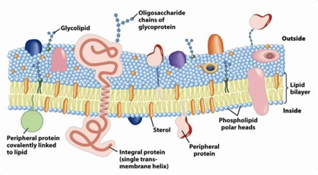
Chapter 5: Cell Membrane Structure – The Fluid Mosaic Model
Understanding cell membrane structure is absolutely crucial for AP Biology success because membrane function underlies virtually every cellular process. The fluid mosaic model, proposed by Singer and Nicolson in 1972, revolutionized our understanding of how membranes work.
Phospholipid Bilayer: The Foundation
Phospholipids are amphipathic molecules – they have both hydrophilic (water-loving) heads and hydrophobic (water-fearing) tails. In an aqueous environment, they spontaneously arrange into a bilayer with hydrophilic heads facing outward and hydrophobic tails pointing inward.
This arrangement creates a selectively permeable barrier. Small, nonpolar molecules like oxygen and carbon dioxide can slip between phospholipids, while larger or polar molecules need assistance.
Membrane Proteins: The Workers
Membrane proteins give each cell type its unique identity and functionality. There are two main categories:
Integral Proteins: These span the entire membrane and often form channels or carriers for transport. Many have hydrophobic regions that interact with the phospholipid tails and hydrophilic regions that extend into the aqueous environments on both sides.
Peripheral Proteins: These associate with the membrane surface and often play roles in cell signaling or maintaining membrane shape.
Cholesterol: The Fluidity Regulator
Cholesterol molecules wedge between phospholipids and regulate membrane fluidity. At high temperatures, cholesterol restrains phospholipid movement, preventing the membrane from becoming too fluid. At low temperatures, it prevents phospholipids from packing too tightly, maintaining necessary fluidity.
Real-World Connection: This is why cold-water fish have different membrane compositions than tropical fish. Arctic fish have more unsaturated fatty acids in their membranes to maintain fluidity in cold water, while tropical fish have more saturated fatty acids to prevent their membranes from becoming too fluid in warm water.
Study Tip: Remember membrane fluidity with the “butter analogy”: saturated fats are like butter (solid at room temperature), while unsaturated fats are like olive oil (liquid at room temperature). More unsaturated fatty acids = more fluid membrane.
Carbohydrates: The Identity Markers
Carbohydrates attached to proteins (glycoproteins) and lipids (glycolipids) on the extracellular surface create the glycocalyx – a carbohydrate coat that serves as cellular identification tags. This is how your immune system recognizes “self” versus “foreign” cells.
Key Takeaways:
- The fluid mosaic model describes membranes as dynamic, flexible structures
- Phospholipid arrangement creates selective permeability
- Membrane proteins determine specific cellular functions
- Cholesterol regulates membrane fluidity
- Carbohydrates serve as cellular identification markers
Chapter 6: Membrane Transport – How Cells Control Their Environment
One of the most heavily tested topics in AP Biology Unit 2 is membrane transport. Understanding these mechanisms is essential because they explain how cells maintain homeostasis, respond to their environment, and carry out essential functions.
Passive Transport: Going with the Flow
Passive transport doesn’t require energy because molecules move down their concentration gradients – from areas of high concentration to areas of low concentration.
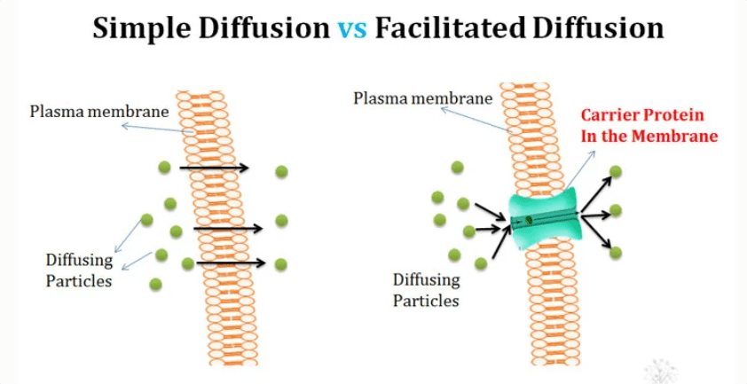
Simple Diffusion: Small, nonpolar molecules like O₂, CO₂, and N₂ can pass directly through the phospholipid bilayer. This process continues until equilibrium is reached.
Facilitated Diffusion: Larger or polar molecules need help from transport proteins. There are two types:
- Channel proteins: Form pores that allow specific ions or molecules to pass through
- Carrier proteins: Bind to specific molecules and change shape to transport them across the membrane
Osmosis: The diffusion of water across a selectively permeable membrane. Water moves from areas of lower solute concentration to areas of higher solute concentration.
Study Tip: Remember osmosis direction with this phrase: “Water follows salt.” Water moves toward the area with more dissolved particles (higher solute concentration).
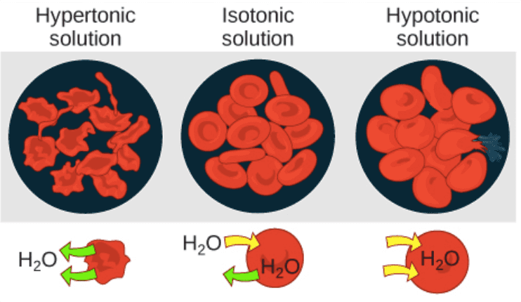
Tonicity: Understanding Water Movement
Tonicity describes the relative solute concentration of solutions separated by a selectively permeable membrane:
Isotonic: Equal solute concentrations on both sides; no net water movement
Hypotonic: Lower solute concentration; water moves into the cell
Hypertonic: Higher solute concentration; water moves out of the cell
Common Mistake Alert: Students often confuse tonicity with concentration. Remember: tonicity describes the solution’s effect on the cell, not just the concentration of solutes.
Active Transport: Working Against the Gradient
Active transport requires energy (usually ATP) to move substances against their concentration gradients – from low to high concentration.
Primary Active Transport: Uses ATP directly. The sodium-potassium pump is the classic example, moving 3 Na⁺ out and 2 K⁺ in against their gradients.
Secondary Active Transport: Uses the energy stored in electrochemical gradients created by primary active transport. For example, glucose is often transported into cells using the sodium gradient established by the Na⁺/K⁺ pump.
Bulk Transport: Moving Large Quantities
When cells need to transport large molecules or large quantities of materials, they use bulk transport processes:
Endocytosis: Bringing materials into the cell
- Phagocytosis: “Cell eating” – engulfing large particles
- Pinocytosis: “Cell drinking” – taking in fluid and dissolved substances
- Receptor-mediated endocytosis: Specific uptake of materials that bind to membrane receptors
Exocytosis: Secreting materials from the cell by fusing vesicles with the cell membrane
Real-World Connection: Understanding endocytosis is crucial for modern medicine. Many drugs are delivered using nanoparticles designed to be taken up by specific cell types through receptor-mediated endocytosis.
Key Takeaways:
- Passive transport moves substances down concentration gradients without energy
- Active transport requires energy to move substances against gradients
- Osmosis and tonicity explain water movement across membranes
- Bulk transport handles large molecules and quantities of materials
Chapter 7: Cell Communication and Signal Transduction
Cells don’t exist in isolation – they constantly communicate with each other through sophisticated signaling mechanisms. This communication allows multicellular organisms to coordinate activities, respond to environmental changes, and maintain homeostasis.
Signal Transduction Overview
Signal transduction involves three main steps:
- Reception: A signaling molecule binds to a receptor protein
- Transduction: The binding triggers a series of molecular changes
- Response: The cell responds with a specific activity
Types of Cell Signaling
Autocrine Signaling: Cells respond to signals they produce themselves
Paracrine Signaling: Cells respond to signals from nearby cells
Endocrine Signaling: Cells respond to hormones carried by the circulatory system
Direct Contact: Cells communicate through direct physical contact
Receptor Types and Locations
Cell Surface Receptors: Located in the cell membrane, these bind to hydrophilic signaling molecules that can’t cross the membrane. Examples include:
- G protein-coupled receptors (GPCRs)
- Receptor tyrosine kinases (RTKs)
- Ion channel receptors
Intracellular Receptors: Located inside the cell, these bind to hydrophobic signaling molecules that can cross the membrane. Examples include steroid hormone receptors.
Study Tip: Remember receptor location by molecule type: “Hydrophilic signals hit surface receptors, hydrophobic signals hit internal receptors.”
Signal Amplification
One of the most important concepts in signal transduction is amplification. A single signaling molecule can trigger a cascade that produces thousands of product molecules, allowing cells to respond dramatically to weak signals.
Real-World Connection: Signal amplification explains how trace amounts of hormones can have dramatic effects. One adrenaline molecule can ultimately trigger the breakdown of millions of glucose molecules, providing rapid energy during fight-or-flight responses.
Key Takeaways:
- Signal transduction involves reception, transduction, and response
- Different signaling types allow for local and long-distance communication
- Receptor location depends on signal molecule properties
- Signal amplification allows dramatic responses to weak signals
Chapter 8: Energy and Enzymes in Cellular Processes
Understanding energy flow and enzymatic reactions is fundamental to grasping all cellular processes. This knowledge connects Unit 2 to upcoming units on cellular respiration and photosynthesis.
Laws of Thermodynamics in Biology
First Law: Energy cannot be created or destroyed, only converted from one form to another. In biological systems, this means the total energy in any reaction remains constant.
Second Law: Every energy transfer increases the entropy (disorder) of the universe. This explains why cellular processes require continuous energy input to maintain organization.
Free Energy and Spontaneous Reactions
Gibbs free energy (ΔG) determines whether a reaction will occur spontaneously:
- ΔG < 0: Exergonic reaction; releases energy; occurs spontaneously
- ΔG > 0: Endergonic reaction; requires energy input; doesn’t occur spontaneously
- ΔG = 0: Reaction is at equilibrium
ATP: The Universal Energy Currency
Adenosine triphosphate (ATP) is the primary energy currency in all living cells. Its structure – adenine, ribose, and three phosphate groups – makes it perfect for this role.
Why ATP Works:
- High-energy phosphate bonds store significant energy
- ATP hydrolysis releases energy for cellular work
- ATP can be quickly regenerated through cellular respiration
ATP Hydrolysis: ATP + H₂O → ADP + Pi + energy
Enzymes: Biological Catalysts
Enzymes are proteins that catalyze biochemical reactions by lowering activation energy. They don’t change the overall energy change of a reaction, but they make reactions occur faster by providing alternative reaction pathways.
Enzyme Structure and Function:
- Active site: The region where substrate binds and catalysis occurs
- Induced fit: The enzyme changes shape slightly when substrate binds
- Enzyme-substrate complex: The temporary association between enzyme and substrate
Factors Affecting Enzyme Activity:
- Temperature: Higher temperatures increase reaction rates until proteins denature
- pH: Each enzyme has an optimal pH range
- Substrate concentration: Higher concentrations increase activity until saturation
- Inhibitors: Competitive and noncompetitive inhibitors reduce activity
Common Mistake Alert: Students often think enzymes provide energy for reactions. Remember: enzymes only lower activation energy; they don’t provide the energy needed for endergonic reactions.
Key Takeaways:
- Thermodynamic laws govern all cellular energy transformations
- Free energy determines reaction spontaneity
- ATP serves as the universal energy currency
- Enzymes catalyze reactions by lowering activation energy
Practice Questions and Exam Strategies
Now let’s put your knowledge to the test with practice questions that mirror the format and difficulty of actual AP Biology exams.
Multiple Choice Questions
Question 1: Which of the following best explains why cells are typically small?
A) Small cells have less DNA to replicate
B) Small cells require less energy to maintain
C) Small cells have a more favorable surface area-to-volume ratio for material exchange
D) Small cells are less likely to be damaged by environmental factors
Answer: C
Explanation: As cells increase in size, volume increases faster than surface area. Since material exchange occurs across the cell surface, larger cells become inefficient at exchanging materials with their environment.
Question 2: A student observes cells under a microscope and notes the presence of chloroplasts, a large central vacuole, and a cell wall. These cells are most likely:
A) Animal cells
B) Bacterial cells
C) Plant cells
D) Fungal cells
Answer: C
Explanation: The combination of chloroplasts, large central vacuole, and cell wall is characteristic of plant cells. While fungal cells have cell walls, they lack chloroplasts and large central vacuoles.
Question 3: Which of the following processes requires the direct input of ATP?
A) Simple diffusion of oxygen across a cell membrane
B) Facilitated diffusion of glucose through a protein channel
C) Osmosis of water through aquaporins
D) Active transport of sodium ions against their concentration gradient
Answer: D
Explanation: Active transport moves substances against their concentration gradients and requires energy input, typically from ATP hydrolysis.
Free Response Practice
Question: Describe the structure and function of the cell membrane, including how its components contribute to selective permeability.
Sample Answer Framework:
- Structure: Fluid mosaic model with phospholipid bilayer, proteins, cholesterol, and carbohydrates
- Selective permeability: Different molecules cross at different rates based on size, polarity, and charge
- Specific examples: O₂ and CO₂ cross easily; glucose requires facilitated diffusion; ions need channels or active transport
- Regulation: Cholesterol affects fluidity; proteins provide specificity
Data Analysis Practice
[INSERT GRAPH: Line graph showing enzyme activity vs. temperature for three different enzymes, with peaks at different temperatures]
Question: The graph above shows enzyme activity versus temperature for three different enzymes. Explain the shape of these curves and predict which enzyme might be found in thermophilic bacteria.
Analysis Points:
- Initial increase due to higher kinetic energy
- Peak represents optimal temperature
- Decline due to protein denaturation
- Enzyme C likely from thermophiles due to higher optimal temperature
Quick Check Knowledge Boxes:
Quick Check 1: Can you explain why mitochondria and chloroplasts have their own DNA?
Answer: They evolved from endosymbiotic bacteria that were engulfed by early eukaryotic cells.
Quick Check 2: What happens to a plant cell in a hypertonic solution?
Answer: Water leaves the cell, causing the cell membrane to pull away from the cell wall (plasmolysis).
Quick Check 3: Why can’t glucose cross the cell membrane without assistance?
Answer: Glucose is polar and too large to pass between phospholipids in the membrane.
Quick Check 4: What’s the difference between active and passive transport?
Answer: Passive transport moves substances down gradients without energy; active transport moves substances against gradients using energy.
Quick Check 5: How do enzymes speed up reactions?
Answer: They lower activation energy by providing alternative reaction pathways.
Common Exam Mistakes and How to Avoid Them
Learning from common mistakes can significantly improve your AP Biology performance. Here are the most frequent errors students make in Unit 2 and strategies to avoid them.
Mistake 1: Confusing Concentration and Tonicity
The Error: Students often say “hypertonic solution has high concentration” without specifying concentration of what.
The Fix: Always specify what you’re comparing. A solution is hypertonic relative to the cell if it has a higher solute concentration than the cell’s interior.
Mistake 2: Misunderstanding Enzyme Function
The Error: Thinking enzymes provide energy for reactions or change the equilibrium of reactions.
The Fix: Remember that enzymes only affect reaction rate by lowering activation energy. They don’t change ΔG or provide energy.
Mistake 3: Oversimplifying Membrane Transport
The Error: Saying “large molecules can’t cross membranes” without considering transport proteins.
The Fix: Specify the type of transport. Large molecules can cross membranes through facilitated diffusion, active transport, or bulk transport.
Mistake 4: Mixing Up Organelle Functions
The Error: Attributing the wrong function to organelles, especially confusing ER types.
The Fix: Create a detailed organelle function chart and review it regularly.
Connecting to Other AP Biology Units
Unit 2 provides the foundation for understanding:
- Unit 3 (Cellular Energetics): Membrane transport is essential for cellular respiration and photosynthesis
- Unit 4 (Cell Communication and Cycle): Signal transduction mechanisms control cell division
- Unit 5 (Heredity): Nuclear structure relates to DNA replication and gene expression
- Unit 6 (Gene Expression): Organelle cooperation is crucial for protein synthesis
- Unit 7 (Natural Selection): Cellular adaptations drive evolutionary change
- Unit 8 (Ecology): Cellular processes scale up to ecosystem functions
Final Thoughts
Mastering Unit 2: Cells requires understanding both structural details and functional relationships. The concepts you’ve learned here – from membrane transport to signal transduction – appear throughout the AP Biology course and exam.
Remember, cellular biology isn’t just about memorizing organelle functions – it’s about understanding how life works at its most fundamental level. When you truly grasp these concepts, the rest of AP Biology becomes much clearer and more interesting.
The investment you make in mastering Unit 2 will pay dividends throughout your AP Biology journey and beyond. Whether you pursue medicine, research, environmental science, or any biology-related field, the cellular foundations you build now will serve you well.
Keep practicing, stay curious, and remember that every expert was once a beginner. You’ve got this!
Also Read –
1 thought on “AP Biology Unit 2: Cells – Complete Study Guide with Practice Questions & Exam Tips”