
Have you ever wondered why you automatically breathe faster when you run up the stairs? Or why mountaineers need oxygen tanks at high altitudes? The answer lies in one of the most elegant systems in your body-the Gas Exchange in Humans. Every single cell in your body is constantly demanding oxygen and producing carbon dioxide, and your respiratory system works tirelessly, every second of every day, to meet these demands. Let’s dive deep into this fascinating topic that’s crucial for your IGCSE Biology 0610 exam!
Understanding the Big Picture: Why Gas Exchange Matters
Before we jump into the technical details, let’s understand why this topic is so important. Your body is like a bustling city with trillions of cells, each acting like a tiny factory. These factories need fuel (glucose) and oxygen to produce energy through aerobic respiration. The waste product? Carbon dioxide, which needs to be removed quickly because it’s toxic in high concentrations.
The gas exchange system is essentially your body’s delivery and waste removal service, working 24/7 without you even thinking about it. You take about 20,000 breaths every day-that’s around 14 breaths per minute when you’re resting. Pretty incredible, right?
The Gas Exchange System: Your Body’s Highway Network
The Respiratory System Components
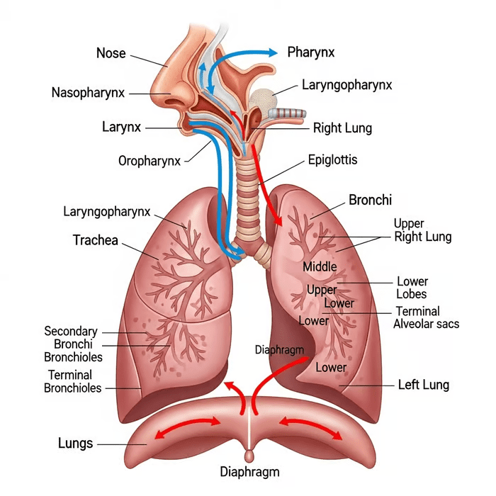
Let’s start our journey with the air you breathe in through your nose or mouth. Think of the respiratory system as a branching tree turned upside down:
The Upper Respiratory Tract:
- Nose and Nasal Cavity: Your nose isn’t just for smelling! The nasal cavity warms, moistens, and filters the air. Tiny hairs called cilia trap dust particles and bacteria, while mucus catches smaller particles. The rich blood supply in the nasal lining warms the air to body temperature.
- Pharynx and Larynx: The pharynx (throat) is a shared pathway for both air and food. The larynx (voice box) contains your vocal cords and has a special flap called the epiglottis that closes over the windpipe when you swallow, preventing food from entering your airways.
The Lower Respiratory Tract:
- Trachea (Windpipe): This is a tube about 12 cm long and 2 cm wide, reinforced with C-shaped rings of cartilage. These rings keep the trachea open at all times-try feeling them at the front of your neck! The incomplete rings (C-shaped, not full circles) allow your esophagus behind the trachea to expand when you swallow food.
- Bronchi: The trachea splits into two bronchi (singular: bronchus), one leading to each lung. They have a similar structure to the trachea with cartilage rings for support.
- Bronchioles: The bronchi branch into smaller and smaller tubes called bronchioles. These don’t have cartilage rings but have smooth muscle in their walls, which can contract or relax to control airflow.
- Alveoli: At the end of the smallest bronchioles are tiny air sacs called alveoli (singular: alveolus). This is where the magic happens! Your lungs contain about 300-500 million alveoli, providing a massive surface area of about 70 square meters-roughly the size of a tennis court packed inside your chest!
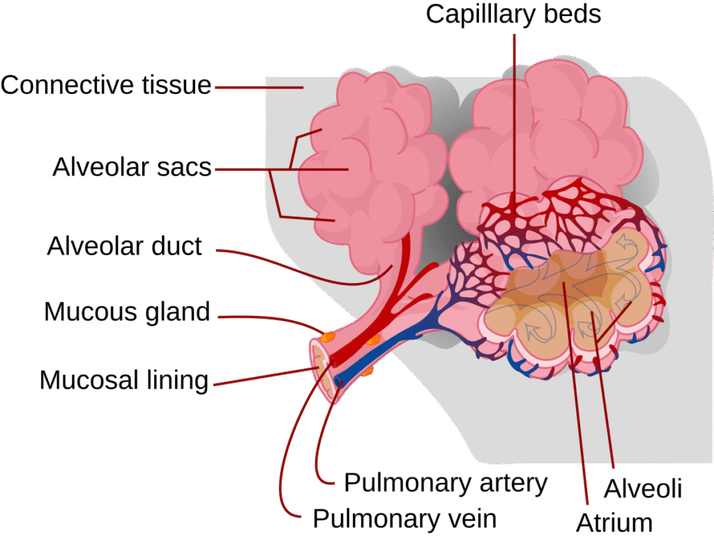
The Lungs: Your Body’s Gas Exchange Powerhouses
Your two lungs sit protected inside your ribcage, with the heart nestled between them. The right lung has three lobes, while the left has only two (making room for your heart). Each lung is surrounded by a double membrane called the pleura, with fluid between the layers that reduces friction during breathing.
Key Features of Lung Structure:
- Spongy and elastic tissue
- Rich blood supply from the pulmonary circulation
- Protected by the ribcage and sternum
- Separated from abdominal organs by the diaphragm
The Stars of Gas Exchange: Alveoli Structure and Function
If the respiratory system were a movie, alveoli would be the main characters. These microscopic air sacs are perfectly designed for efficient gas exchange. Let’s explore why they’re so good at their job:
Alveolar Structure: Design Perfection
Walls Just One Cell Thick:
The alveolar walls consist of a single layer of flattened epithelial cells. This minimal thickness means gases only have to travel a tiny distance-about 0.5 micrometers (that’s 0.0005 mm)! This short diffusion pathway allows rapid gas exchange.
Surrounded by Capillaries:
Each alveolus is wrapped in a dense network of capillaries (tiny blood vessels). The capillary walls are also just one cell thick. Between the alveolar wall and capillary wall, there’s a thin layer of moisture where gases dissolve.
Moist Surface:
The inside of alveoli is covered with a thin film of moisture. Oxygen must first dissolve in this liquid before it can diffuse into the blood. This moisture is essential for gas exchange.
Elastic Walls:
Alveolar walls contain elastic fibers that stretch when you breathe in and recoil when you breathe out, helping to push air out of the lungs.
Rich Blood Supply:
The pulmonary artery brings deoxygenated blood from the heart to the lungs, splitting into millions of capillaries around the alveoli. After gas exchange, the oxygenated blood travels back to the heart via the pulmonary veins.
Why Alveoli Are So Efficient: The Adaptations
Let’s summarize the key adaptations that make alveoli perfect for gas exchange:
| Adaptation | How It Helps Gas Exchange |
|---|---|
| Large surface area (millions of alveoli) | More space for gas exchange to occur simultaneously |
| Thin walls (one cell thick) | Short diffusion distance for gases (Fick’s Law) |
| Moist lining | Gases dissolve before diffusing |
| Rich blood supply | Maintains concentration gradient by constantly bringing new blood |
| Good ventilation | Maintains concentration gradient by refreshing air supply |
| Elastic tissue | Helps with breathing out and prevents alveolar collapse |
Memory Tip: Use the mnemonic “SLAVE” for alveolar adaptations:
- Surface area (large)
- Lining (moist)
- Alveolar walls (thin)
- Ventilation (good)
- Exchange surface (rich blood supply)
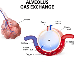
The Process of Gas Exchange: Following the Oxygen
Now let’s trace the journey of oxygen from the air into your bloodstream, and carbon dioxide making the reverse journey:
Step 1: Ventilation (Breathing In)
When you inhale, fresh air rich in oxygen (about 21% oxygen, 0.04% carbon dioxide) enters the alveoli. The concentration of oxygen in the alveolar air is high.
Step 2: Oxygen Diffusion
Oxygen dissolves in the moisture lining the alveolus and then diffuses across the alveolar wall and capillary wall into the blood plasma. From there, it diffuses into red blood cells where it binds with hemoglobin to form oxyhemoglobin:
Oxygen + Hemoglobin → Oxyhemoglobin
Each hemoglobin molecule can carry up to four oxygen molecules. This is crucial because oxygen isn’t very soluble in blood plasma alone.
Why does oxygen move into the blood? Because of diffusion! Oxygen moves from an area of high concentration (alveolar air) to an area of low concentration (deoxygenated blood in capillaries). This is called a concentration gradient.
Step 3: Carbon Dioxide Diffusion
Meanwhile, carbon dioxide in the blood (produced by respiring cells throughout your body) is at a high concentration. It diffuses from the blood into the alveolar air where the concentration is lower. Carbon dioxide is transported in blood in three ways:
- About 7% dissolved in plasma
- About 23% bound to hemoglobin
- About 70% as hydrogen carbonate ions (HCO₃⁻) in plasma
Step 4: Breathing Out
When you exhale, the air you breathe out has less oxygen (about 16%) and more carbon dioxide (about 4%) than the air you breathed in.
Breathing Mechanisms: How Air Moves In and Out
Many students confuse breathing with respiration, so let’s clear this up right now:
Breathing (Ventilation): The mechanical process of moving air in and out of the lungs
Respiration: The chemical process of releasing energy from glucose in cells
Breathing involves two phases: inspiration (breathing in) and expiration (breathing out).
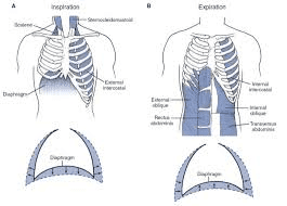
Inspiration: The Active Process
Breathing in requires energy and muscular effort:
Step-by-Step Process:
- Brain signals: Your brain (specifically the medulla oblongata) sends signals to breathing muscles
- Diaphragm contracts: This dome-shaped muscle flattens and moves downward
- External intercostal muscles contract: These muscles between your ribs pull the ribcage upward and outward
- Volume increases: The thoracic cavity (chest cavity) volume increases
- Pressure decreases: As volume increases, air pressure inside the lungs decreases below atmospheric pressure
- Air rushes in: Air flows from high pressure (outside) to low pressure (inside lungs)
Think of it like pulling a syringe plunger-the volume inside increases, pressure drops, and air rushes in!
Expiration: Usually Passive
Breathing out at rest doesn’t normally require energy:
Step-by-Step Process:
- Muscles relax: The diaphragm and external intercostal muscles relax
- Diaphragm moves up: It returns to its dome shape
- Ribcage moves down and in: Due to gravity and elastic recoil
- Volume decreases: Thoracic cavity volume decreases
- Pressure increases: Air pressure inside lungs increases above atmospheric pressure
- Air flows out: Air moves from high pressure (lungs) to low pressure (outside)
During Exercise: Expiration becomes active. Internal intercostal muscles contract to pull the ribcage down more forcefully, and abdominal muscles contract to push the diaphragm up higher, expelling more air.
The Role of the Pleural Membranes
The pleural membranes and pleural fluid between them are crucial for breathing. The fluid creates surface tension that helps the lungs stick to the inside of the ribcage and allows smooth movement. Without this, breathing would be difficult and painful.
Composition of Inspired and Expired Air: The Numbers Matter
This is a favorite exam topic, so memorize these values!
| Gas | Inspired Air (%) | Expired Air (%) | Change |
|---|---|---|---|
| Oxygen | 21 | 16 | Decreased by 5% |
| Carbon dioxide | 0.04 | 4 | Increased by 100 times! |
| Nitrogen | 78 | 78 | No change |
| Water vapor | Variable | Saturated | Increased |
| Temperature | Variable | Body temperature (37°C) | Usually increased |
Key Points to Remember:
- Oxygen decreases because it’s absorbed into the blood
- Carbon dioxide increases dramatically because it’s expelled from the blood
- Nitrogen doesn’t change because it’s not used in the body
- Expired air is warmer and more humid (saturated with water vapor)
- Expired air still contains 16% oxygen-that’s why mouth-to-mouth resuscitation works!
Common Breathing Experiments: Practical Biology
The Limewater Test
Purpose: To detect carbon dioxide
Method:
- Breathe out through a straw into clear limewater
- The limewater turns cloudy/milky
- This proves expired air contains more CO₂ than inspired air
Chemical Reaction:
Carbon dioxide + Calcium hydroxide → Calcium carbonate (white precipitate) + Water
CO₂ + Ca(OH)₂ → CaCO₃ + H₂O
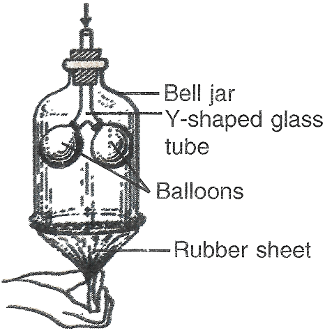
The Bell Jar Model
This is a classic demonstration of breathing mechanics:
Components:
- Bell jar (represents ribcage and chest wall)
- Rubber sheet at bottom (represents diaphragm)
- Y-shaped tube (represents trachea and bronchi)
- Balloons (represent lungs)
How It Works:
- Pull the rubber sheet down → volume increases, pressure decreases, balloons inflate (inspiration)
- Push rubber sheet up → volume decreases, pressure increases, balloons deflate (expiration)
Limitations: This model is simplified-real lungs are elastic, the ribcage moves, and breathing is controlled by the brain, not manual manipulation.
Factors Affecting Breathing Rate
Your breathing isn’t constant-it changes based on your body’s needs:
Exercise: Muscles need more oxygen and produce more CO₂, so breathing rate and depth increase (from about 15 to 40-60 breaths per minute)
Carbon Dioxide Concentration: The primary stimulus for breathing! Chemoreceptors in the brain detect CO₂ levels in blood. High CO₂ triggers faster, deeper breathing.
Oxygen Levels: At high altitudes or in oxygen-poor environments, low oxygen levels also stimulate breathing rate.
Emotions: Stress, fear, or excitement can increase breathing rate through nervous system activation.
Temperature: Hot conditions can increase breathing rate; cold can decrease it.
Health Conditions: Fever, lung diseases, and other illnesses affect breathing.
Health and Disease: When Gas Exchange Goes Wrong
Understanding normal gas exchange helps us understand respiratory diseases:
Asthma
- Airways become inflamed and narrow
- Smooth muscle in bronchioles contracts
- Mucus production increases
- Results in difficulty breathing, wheezing, coughing
- Treatment: Inhalers (bronchodilators to relax muscles, steroids to reduce inflammation)
Emphysema
- Alveolar walls break down, reducing surface area
- Often caused by smoking
- Results in reduced gas exchange efficiency
- Breathing becomes difficult, especially during exercise
Pneumonia
- Alveoli fill with fluid and pus
- Reduces gas exchange surface area
- Caused by bacterial or viral infection
Effects of Smoking
Smoking damages the respiratory system in multiple ways:
- Tar coats alveoli, reducing gas exchange
- Carbon monoxide in smoke binds to hemoglobin more strongly than oxygen, reducing oxygen transport
- Irritants damage cilia, reducing the cleaning mechanism
- Increases risk of lung cancer, bronchitis, and emphysema
Quick Revision: Key Points Summary
✓ Gas exchange occurs in the alveoli through diffusion across thin, moist membranes
✓ Alveoli adaptations: Large surface area, thin walls, moist lining, rich blood supply, good ventilation (Remember: SLAVE)
✓ Breathing mechanism: Diaphragm and intercostal muscles change thoracic volume, changing pressure, causing air flow
✓ Inspiration is active (muscles contract), expiration is passive at rest (muscles relax)
✓ Oxygen diffuses from alveoli → blood; Carbon dioxide diffuses from blood → alveoli
✓ Composition changes: Expired air has less O₂ (16% vs 21%), more CO₂ (4% vs 0.04%), is warmer and more humid
✓ Trachea has C-shaped cartilage rings to keep airways open
✓ Hemoglobin carries oxygen as oxyhemoglobin
✓ CO₂ is the main breathing stimulus, detected by chemoreceptors in the brain
✓ Don’t confuse breathing (ventilation) with respiration (energy release in cells)

Important Equations and Formulas
Formation of Oxyhemoglobin:
Hemoglobin + Oxygen ⇌ Oxyhemoglobin
Hb + 4O₂ ⇌ HbO₈
(This is a reversible reaction)
Limewater Test:
CO₂ + Ca(OH)₂ → CaCO₃ + H₂O
(Calcium hydroxide turns cloudy white)
Link to Aerobic Respiration:
Glucose + Oxygen → Carbon dioxide + Water + Energy (ATP)
C₆H₁₂O₆ + 6O₂ → 6CO₂ + 6H₂O + Energy
The oxygen obtained through gas exchange is used for this cellular respiration process!
Test Yourself: Practice Questions
Question 1: Explain three ways in which alveoli are adapted for efficient gas exchange. (6 marks)
Question 2: Describe the process of inspiration. (4 marks)
Question 3: Complete the table showing the composition of inspired and expired air:
| Gas | Inspired (%) | Expired (%) |
|---|---|---|
| Oxygen | ? | ? |
| Carbon dioxide | ? | ? |
| Nitrogen | ? | ? |
Question 4: Explain why expired air still contains oxygen. (2 marks)
Question 5: The diagram shows a bell jar model of breathing. Explain one limitation of this model. (2 marks)
Question 6: State the difference between breathing and respiration. (2 marks)
Question 7: Explain why breathing rate increases during exercise. (3 marks)
Question 8: Describe how oxygen moves from the alveolar air into the red blood cells. (4 marks)
Sample Answers (Check Your Work!)
Answer 1:
- Large surface area (millions of alveoli) provides more space for gas exchange
- Thin walls (one cell thick) create a short diffusion distance for gases
- Moist lining allows gases to dissolve before diffusing
- Rich blood supply maintains concentration gradient by constantly removing oxygen and bringing carbon dioxide
- Good ventilation maintains concentration gradient by constantly refreshing air supply
- Elastic tissue allows alveoli to stretch and recoil during breathing
Answer 2:
- External intercostal muscles contract
- Diaphragm contracts and flattens
- Volume of thoracic cavity increases
- Pressure inside lungs decreases below atmospheric pressure
- Air moves into the lungs from higher to lower pressure
Answer 3:
Oxygen: 21% → 16%
Carbon dioxide: 0.04% → 4%
Nitrogen: 78% → 78%
Common Mistakes to Avoid
Saying “oxygen is released during gas exchange” → Oxygen is ABSORBED, carbon dioxide is RELEASED
Confusing breathing with respiration → Breathing is physical air movement; respiration is chemical energy release in cells
Saying the diaphragm moves up during inspiration → It moves DOWN and flattens
Thinking all expired air components change → Nitrogen stays the same!
Saying cartilage rings are complete circles → They’re C-shaped to allow the esophagus to expand
Forgetting that expiration is passive at rest → It becomes active only during exercise or forced breathing
Saying gases move by active transport → Gas exchange occurs by DIFFUSION (passive process)
Thinking alveoli have thick walls → They’re ONE CELL THICK for efficient diffusion
Study Tips for Exam Success
1. Practice Drawing Diagrams: You might need to label the respiratory system or draw an alveolus. Practice regularly!
2. Learn the Numbers: Memorize the percentages for inspired and expired air composition—these appear in almost every exam.
3. Understand Movement: Make sure you know which way the diaphragm and ribs move during inspiration and expiration. Use your own body to feel it!
4. Connect Topics: Link gas exchange to respiration (Topic 12) and transport in humans (Topic 10)—they work together as a system.
5. Use Past Papers: Practice questions from past Cambridge papers specifically on gas exchange. Look for patterns in how questions are asked.
6. Master Command Words:
- Describe = say what happens
- Explain = say what happens AND why
- State = short answer, no explanation needed
7. Learn Correct Terminology: Use scientific terms like “concentration gradient,” “diffusion,” “thoracic cavity,” not everyday language.
Real-Life Applications: Why This Matters
Understanding gas exchange helps you appreciate:
- Why athletes train at high altitude: Lower oxygen forces the body to produce more red blood cells, improving performance at sea level
- How ventilators work: They mechanically change thoracic volume for patients who can’t breathe independently
- Why exercise makes you breathless: Your muscles use oxygen faster than you can supply it, creating “oxygen debt”
- How mouth-to-mouth resuscitation saves lives: Your expired air (16% oxygen) still has enough oxygen to help someone who isn’t breathing
- Why scuba divers need training: Understanding pressure changes and gas exchange prevents dangerous conditions like “the bends”
- Why premature babies need special care: Their alveoli may lack surfactant, making breathing difficult
Final Motivation
Gas exchange is one of the most beautifully designed systems in your body. Every breath you take involves millions of alveoli working simultaneously, maintaining the delicate balance that keeps you alive. The fact that this happens automatically, about 20,000 times a day without conscious thought, is truly remarkable.
When you’re studying late at night and feeling tired, remember that your respiratory system is working perfectly to supply your brain with oxygen for all that learning. Your understanding of this topic isn’t just about passing an exam-it’s about understanding the incredible machine that is your own body.
You’ve got this! Keep breathing, keep learning, and success will follow. The IGCSE Biology 0610 exam is just one milestone on your educational journey, and with thorough understanding of topics like gas exchange, you’re building a foundation for future success in science.
Remember: Every expert was once a beginner. Every respiratory physiologist once sat where you’re sitting, learning these exact same concepts. Your future is as bright as your determination.
Happy studying, and breathe easy-you’re going to ace this!
Recommended –
1 thought on “Gas Exchange in Humans | IGCSE Biology Topic 11 Complete Guide”