
Transport in Animals: The Ultimate IGCSE Biology Guide
Have you ever wondered how oxygen from your lungs reaches your toes? Or how the sandwich you ate at lunch provides energy to your brain cells? The answer lies in one of nature’s most incredible engineering marvels – your circulatory system!
Think of your body as a bustling city. Just like a city needs roads, trains, and delivery trucks to transport goods and people, your body needs a sophisticated transport system to move nutrients, oxygen, waste products, and chemical messages. In this comprehensive guide, we’ll explore how animals – including humans – have evolved remarkable transport systems to keep every cell alive and functioning.
Whether you’re preparing for your IGCSE Biology exam or simply curious about how your body works, this guide will take you on a fascinating journey through blood vessels, beating hearts, and life-sustaining circulation. Let’s dive in!
Why Do Animals Need Transport Systems?
Before we explore the “how,” let’s understand the “why.”
Single-celled organisms like amoeba can survive through simple diffusion – substances move directly across their cell membrane. But here’s the problem: as organisms get bigger, their surface area to volume ratio decreases dramatically.
What does this mean?
- Large animals have many cells deep inside their bodies
- These cells are too far from the surface for diffusion to work efficiently
- Diffusion alone would be too slow to meet cellular demands
This is where specialized transport systems come in!
Animals need transport systems to:
- Deliver oxygen and nutrients to all body cells
- Remove carbon dioxide and other metabolic waste products
- Distribute hormones (chemical messengers)
- Transport antibodies and white blood cells for immune defense
- Maintain body temperature through heat distribution
The Human Circulatory System: An Overview
The human circulatory system (also called the cardiovascular system) consists of three main components working together like a perfectly choreographed dance:
1. The Heart – The Pump
A muscular organ about the size of your fist that beats approximately 100,000 times per day, pumping around 7,000 litres of blood!
2. Blood Vessels – The Network
An extensive system of tubes (arteries, veins, and capillaries) stretching about 100,000 kilometers – enough to circle the Earth twice!
3. Blood – The Transport Medium
A specialized liquid tissue that carries everything your cells need and takes away what they don’t.
The Heart: Your Body’s Tireless Pump
Structure of the Heart
The human heart is divided into four chambers – think of it as a two-story house with two rooms on each floor:
Upper Chambers (Atria – singular: atrium)
- Right atrium: Receives deoxygenated blood from the body via the vena cava
- Left atrium: Receives oxygenated blood from the lungs via the pulmonary vein
Lower Chambers (Ventricles)
- Right ventricle: Pumps deoxygenated blood to the lungs via the pulmonary artery
- Left ventricle: Pumps oxygenated blood to the body via the aorta
Key Observation: The left ventricle has the thickest muscular wall because it needs to pump blood all around the entire body, whereas the right ventricle only pumps to the nearby lungs.
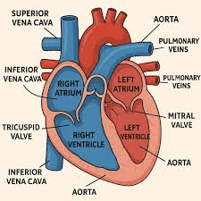
Valves: The One-Way Traffic Controllers
Hearts contain four valves that ensure blood flows in only one direction – preventing backflow:
- Tricuspid valve: Between right atrium and right ventricle (has 3 flaps)
- Bicuspid valve (Mitral valve): Between left atrium and left ventricle (has 2 flaps)
- Pulmonary semi-lunar valve: Between right ventricle and pulmonary artery
- Aortic semi-lunar valve: Between left ventricle and aorta
Memory Tip: Think “TRI-cuspid = 3 flaps = RIGHT side” and “BI-cuspid = 2 flaps = LEFT side”
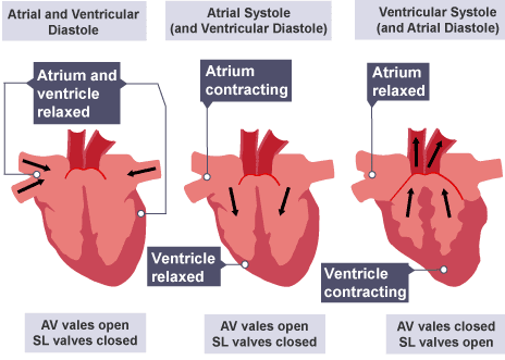
The Cardiac Cycle: How Your Heart Beats
The heart beats in a coordinated rhythm called the cardiac cycle, which has three main phases:
Phase 1: Atrial Systole (Atria Contract)
- Both atria contract simultaneously
- Blood is pushed through open valves into the ventricles
- Ventricles are relaxed and filling with blood
Phase 2: Ventricular Systole (Ventricles Contract)
- Both ventricles contract with great force
- Pressure closes the tricuspid and bicuspid valves (creates “lub” sound)
- Blood is forced out through the pulmonary artery and aorta
- Semi-lunar valves are open
Phase 3: Diastole (Complete Relaxation)
- All chambers relax
- Semi-lunar valves close (creates “dub” sound)
- Blood from the vena cava and pulmonary vein flows into the atria
- The cycle begins again
The familiar “lub-dub” heartbeat sound you hear with a stethoscope is actually the valves closing!
Coronary Heart Disease: When Things Go Wrong
The heart muscle itself needs oxygen and nutrients, supplied by coronary arteries that branch from the aorta.
What happens in coronary heart disease?
- Fatty deposits (plaques) build up in coronary arteries
- This narrows the arteries (a process called atherosclerosis)
- Blood flow to heart muscle is reduced
- Can lead to chest pain (angina) or heart attack (myocardial infarction)
Risk Factors:
- High cholesterol diet (excessive saturated fats)
- Smoking
- Lack of exercise
- High blood pressure
- Genetic factors
- Stress
- Obesity
Prevention Methods:
- Balanced diet low in saturated fats
- Regular exercise
- Avoiding smoking
- Managing stress levels
- Maintaining healthy weight
Blood Vessels: The Highway Network
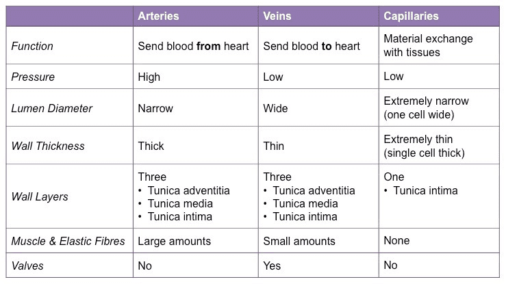
Your body contains three types of blood vessels, each perfectly designed for its specific function:
1. Arteries: The High-Pressure Highways
Structure:
- Thick, muscular walls with elastic tissue
- Small lumen (internal space) relative to wall thickness
- No valves (except where they leave the heart)
Function:
- Carry blood away from the heart
- Transport blood under high pressure
- Muscular walls help maintain blood pressure
- Elastic walls stretch and recoil with each heartbeat (creates pulse)
Special Note: The pulmonary artery is unique – it’s the only artery carrying deoxygenated blood (from heart to lungs).
2. Veins: The Return Journey
Structure:
- Thinner walls with less muscle and elastic tissue
- Larger lumen relative to wall thickness
- Contains valves at intervals
Function:
- Carry blood toward the heart
- Transport blood under low pressure
- Valves prevent backflow of blood
- Surrounded by muscles that squeeze veins, pushing blood forward
How do veins overcome gravity? When you stand up, blood in leg veins must flow upward against gravity. This is achieved through:
- Valves: Stop blood flowing backward
- Muscle action: Contracting leg muscles squeeze veins
- Breathing movements: Changes in chest pressure during breathing help pull blood upward
Special Note: The pulmonary vein is unique – it’s the only vein carrying oxygenated blood (from lungs to heart).
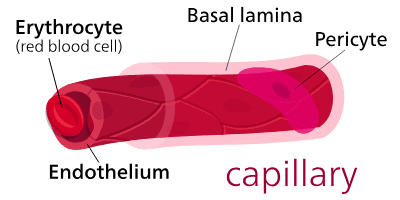
3. Capillaries: The Delivery Network
Structure:
- Extremely thin walls (one cell thick)
- Microscopic diameter (just wide enough for red blood cells to squeeze through single file)
- Form extensive networks in all tissues
Function:
- Site of exchange between blood and tissues
- Thin walls allow rapid diffusion of substances
- Large surface area for maximum exchange
- Short diffusion distance
What gets exchanged at capillaries?
- Into tissues: Oxygen, glucose, amino acids, hormones, antibodies
- Out of tissues: Carbon dioxide, urea, other waste products
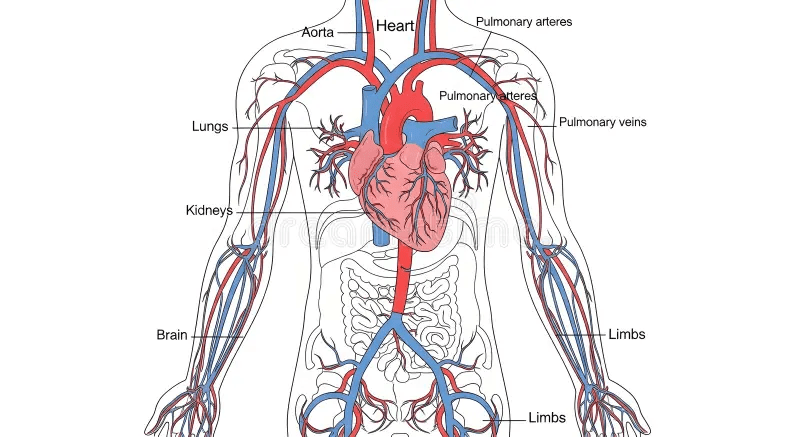
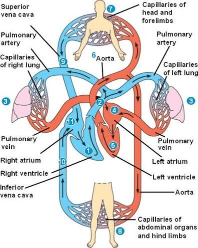
The Double Circulatory System
Humans and other mammals have a double circulatory system – blood passes through the heart twice during one complete circuit. This is much more efficient than a single circulatory system!
Circuit 1: Pulmonary Circulation (Heart → Lungs → Heart)
The Route:
- Right ventricle → Pulmonary artery → Lungs
- Gas exchange occurs in lung capillaries (oxygen in, carbon dioxide out)
- Lungs → Pulmonary vein → Left atrium
Why is this important? Blood pressure drops significantly as blood passes through the lung capillaries. Without a double system, this low-pressure blood would flow very slowly around the body.
Circuit 2: Systemic Circulation (Heart → Body → Heart)
The Route:
- Left ventricle → Aorta → Body organs
- Exchange occurs in tissue capillaries (oxygen and nutrients out, carbon dioxide and waste in)
- Body → Vena cava → Right atrium
The Advantage: Blood returns to the heart to be pumped out again at high pressure, ensuring rapid delivery of oxygen and nutrients to all body cells.
Blood: The Amazing Transport Medium
Blood is a specialized liquid tissue composed of:
1. Plasma (55% of blood volume)
What is it? A pale yellow liquid consisting of 90% water plus dissolved substances.
What does it carry?
- Nutrients: Glucose, amino acids, vitamins, minerals
- Waste products: Urea, carbon dioxide
- Hormones: Chemical messengers
- Proteins: Antibodies, fibrinogen (for clotting)
- Heat: Helps distribute warmth around the body
2. Red Blood Cells (Erythrocytes) – 45% of blood volume
Special Adaptations:
- Biconcave disc shape: Increases surface area for gas exchange
- No nucleus: More space for hemoglobin
- Contains hemoglobin: Red protein that binds oxygen
- Flexible: Can squeeze through narrow capillaries
Function: Transport oxygen from lungs to all body cells
The Chemistry:
- In lungs (high oxygen): Hemoglobin + Oxygen → Oxyhemoglobin (bright red)
- In tissues (low oxygen): Oxyhemoglobin → Hemoglobin + Oxygen (dark red)
Memory Tip: “Red Blood Cells are RED because of Hemoglobin, which carries oxygen to help cells RESPIRE”
3. White Blood Cells (Leukocytes) – Less than 1%
Function: Defend the body against disease
Types:
- Phagocytes: Engulf and digest bacteria and viruses
- Lymphocytes: Produce antibodies that target specific pathogens
Key Features:
- Have a nucleus (unlike red blood cells)
- Can change shape to squeeze out of blood vessels
- Can move independently through tissues
4. Platelets (Thrombocytes) – Less than 1%
What are they? Tiny cell fragments without a nucleus
Function: Blood clotting to prevent blood loss
How Clotting Works:
- Blood vessel is damaged
- Platelets arrive and stick to damaged area
- Platelets release chemicals
- These chemicals convert fibrinogen (soluble protein) → fibrin (insoluble threads)
- Fibrin forms a mesh that traps blood cells
- This forms a clot (scab), sealing the wound
Comparison of Blood Components
| Component | Structure | Function | Percentage |
|---|---|---|---|
| Plasma | Pale yellow liquid | Transport dissolved substances, heat distribution | 55% |
| Red Blood Cells | Biconcave, no nucleus, contains hemoglobin | Oxygen transport | 45% |
| White Blood Cells | Various types, have nucleus | Fight disease | <1% |
| Platelets | Cell fragments, no nucleus | Blood clotting | <1% |
Quick Revision: Key Concepts Summary
✓ Why transport systems? Large animals need them because diffusion is too slow over long distances
✓ Heart structure: Four chambers (2 atria, 2 ventricles), four valves, left ventricle has thickest wall
✓ Cardiac cycle: Atrial systole → Ventricular systole → Diastole
✓ Blood vessels:
- Arteries: Thick walls, high pressure, blood away from heart
- Veins: Thin walls, valves, low pressure, blood toward heart
- Capillaries: One cell thick, site of exchange
✓ Double circulation:
- Pulmonary circuit: Right heart → Lungs → Left heart
- Systemic circuit: Left heart → Body → Right heart
✓ Blood components:
- Plasma: Transport medium
- Red blood cells: Oxygen transport (hemoglobin)
- White blood cells: Defense
- Platelets: Clotting
✓ Coronary heart disease: Fatty deposits narrow coronary arteries, reducing blood flow to heart muscle
Common Mistakes to Avoid
Mistake 1: Saying “all arteries carry oxygenated blood”
Correction: The pulmonary artery carries deoxygenated blood from the heart to the lungs
Mistake 2: Confusing the vena cava with the aorta
Remember: Vena cava brings blood TO the heart; aorta takes blood AWAY from the heart
Mistake 3: Thinking valves are found in arteries
Correction: Valves are only in veins (and in the heart)
Mistake 4: Saying red blood cells carry carbon dioxide
Correction: Red blood cells mainly carry oxygen; carbon dioxide is mostly transported dissolved in plasma
Mistake 5: Forgetting that capillaries are the site of exchange
Remember: Arteries and veins transport; capillaries exchange
Mistake 6: Mixing up atria and ventricles
Remember: Atria are on top (think “attic”), ventricles are on the bottom
Important Exam-Style Questions
Question 1: Structure and Function
Q: Explain why the left ventricle has a thicker muscular wall than the right ventricle. (3 marks)
Model Answer:
- The left ventricle pumps blood all around the body (1 mark)
- The right ventricle only pumps blood to the lungs, which are nearby (1 mark)
- Therefore, the left ventricle needs thicker muscle to generate higher pressure (1 mark)
Question 2: Blood Vessels
Q: Complete the table comparing arteries and veins. (4 marks)
| Feature | Artery | Vein |
|---|---|---|
| Wall thickness | Thick | Thin |
| Presence of valves | No (except at heart) | Yes |
| Blood pressure | High | Low |
| Lumen size | Small relative to wall | Large relative to wall |
Question 3: Heart Function
Q: Describe what happens during ventricular systole. (3 marks)
Model Answer:
- Both ventricles contract (1 mark)
- The tricuspid and bicuspid valves close (1 mark)
- Blood is forced out through the pulmonary artery and aorta (1 mark)
Question 4: Blood Components
Q: Explain how red blood cells are adapted for their function. (4 marks)
Model Answer:
- Biconcave disc shape increases surface area for oxygen absorption (1 mark)
- No nucleus provides more space for hemoglobin (1 mark)
- Contains hemoglobin which binds to oxygen (1 mark)
- Flexible so can squeeze through narrow capillaries (1 mark)
Question 5: Double Circulation
Q: Explain the advantage of a double circulatory system. (3 marks)
Model Answer:
- Blood passes through the heart twice in one complete circuit (1 mark)
- Blood pressure drops after passing through lung capillaries (1 mark)
- Blood is returned to the heart and pumped out at high pressure, ensuring rapid delivery of oxygen and nutrients to body tissues (1 mark)
Question 6: Application
Q: A person’s coronary arteries become narrowed due to fatty deposits. Explain why this may cause chest pain during exercise. (3 marks)
Model Answer:
- Coronary arteries supply the heart muscle with oxygen and nutrients (1 mark)
- Narrowed arteries reduce blood flow to heart muscle (1 mark)
- During exercise, the heart needs more oxygen, but narrowed arteries cannot supply enough, causing pain (1 mark)
Memory Tips and Mnemonics
For Blood Flow Through the Heart: “Victor Creates Awesome Lessons”
- Vena cava → Right atrium → Tricuspid valve
- Cardiac (right ventricle) → Pulmonary artery → Lungs
- Alveoli exchange → Pulmonary vein → Left atrium
- Left ventricle → Aorta → Body
For Artery vs. Vein: “Arteries Away, Veins Toward”
- Arteries carry blood Away from the heart
- Veins carry blood Toward the heart (return to heart)
For Valve Names: “Try Being My Assistant”
- Tricuspid valve (right side)
- Bicuspid valve (left side)
- Main arteries have semi-lunar valves
For Blood Components: “Please Red Wine Pills”
- Plasma (liquid part)
- Red blood cells (oxygen carriers)
- White blood cells (warriors against disease)
- Platelets (plug wounds)
Real-Life Connections
Exercise and Your Heart
When you exercise, your muscles need more oxygen and glucose. Your heart responds by:
- Beating faster (increased heart rate)
- Pumping more blood per beat (increased stroke volume)
- This is why you can feel your pulse racing during a run!
Taking Your Pulse
You can feel your pulse at your wrist or neck. What you’re feeling is:
- The surge of blood as your left ventricle contracts
- The elastic artery walls stretching and recoiling
- A typical resting pulse is 60-100 beats per minute
Blood Types and Donation
Your blood type (A, B, AB, or O) affects compatibility for transfusions. This is why blood donation saves lives – but types must match!
High Altitude Adaptation
People living at high altitudes (like in the Himalayas) produce more red blood cells to compensate for lower oxygen levels. This is why athletes sometimes train at altitude!
Key Equations and Formulas Box
Cardiac Output Equation:
Cardiac Output = Heart Rate × Stroke Volume
(volume of blood pumped per minute = beats per minute × volume per beat)
Hemoglobin Chemistry:
In lungs: Hemoglobin + Oxygen → Oxyhemoglobin

In tissues: Oxyhemoglobin → Hemoglobin + Oxygen
Blood Clotting Cascade:
Platelet chemicals trigger:
Fibrinogen (soluble) → Fibrin (insoluble threads) → Blood clot forms
Surface Area to Volume Ratio:
As size increases, SA:V ratio decreases
→ Diffusion becomes inefficient
→ Transport system needed
Keep Going!
Remember, every doctor, surgeon, and biologist started exactly where you are now. The human circulatory system is complex, but you’ve taken the first important steps to mastering it.
Your heart has beaten approximately 3,000 times since you started reading this guide – that’s 3,000 reminders of the incredible biological machinery working tirelessly to keep you alive!
Final Encouragement
Transport in animals isn’t just an exam topic – it’s the story of life itself. Every time your heart beats, you’re experiencing millions of years of evolutionary perfection. Every breath you take, every step you run, every thought you think depends on this remarkable system working flawlessly.
When exam questions seem tough, remember: you’re learning about the system that’s keeping you alive right this second. That’s pretty amazing!
You’ve got this!
Now go ace that IGCSE Biology exam! Your heart believes in you – after all, it’s literally supporting you every step of the way!
Recommended –
1 thought on “IGCSE (Cambridge) Biology Topic 9: Transport in Animals | Your Complete Study Guide”